무릎 및 종아리
이론과 하이라이트 히스토리를 확인 할 수 있어요.
0. 무릎
Patella가 가장 중요한 뼈이다.
Patella 개요 | |
|---|---|
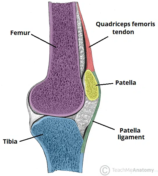
위치 | Femur의 patellofemoral groove 내 |
연결 구조 | 위쪽: Quadriceps tendon / 아래쪽: Patellar ligament |
형태 | 삼각형 모양, 앞면(anterior)과 뒷면(posterior) 구분 |
특징 | 인체 최대의 Sesamoid bone (건 내 삽입된 뼈) quadriceps tendon 내에 위치하여 무릎의 신전 작용 시 지렛대 효과를 증가시킴 |
Patella의 Landmark는 다음과 같다.
구조 | 설명 |
|---|---|
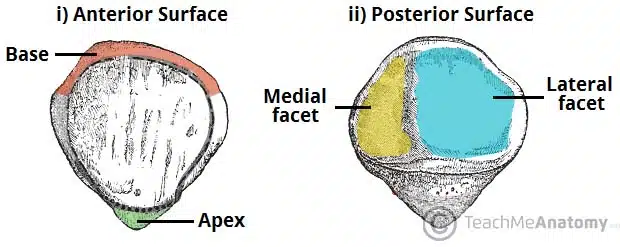
Base | 상단부, Quadriceps tendon 부착 부위 |
Apex | 하단부, Patellar ligament를 통해 Tibial tuberosity와 연결 |
Posterior surface | Femur와 관절면 형성 |
1) 무릎 관절 개요
(1) 유형: 경첩형 활막관절 (hinge-type synovial joint)
(2) 기능: 굴곡(flexion), 신전(extension), (굴곡 상태에서) 내회전/외회전
(3) Tibiofemoral joint: femoral condyles와 tibial condyles 간의 관절. 체중 부하의 주축.
(4) Patellofemoral joint: patella와 femur 전방 간의 관절. quadriceps femoris의 효율적 작용을 도움.
2) 혈관 및 신경
(1) 혈관: genicular branches of femoral a. & popliteal a.
(2) 신경 (Hilton’s law): Femoral n., Tibial n., Common fibular n.
3) 반월상연골 (Meniscus)
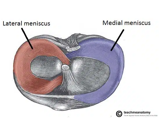
(1) Medial meniscus: medial collateral lig.와 관절낭에 부착. 손상 시 medial lig.와 함께 손상 가능성 높음.
(2) Lateral meniscus: 추가 부착이 없어 더 가동성이 있음.
(3) 기능: 관절면을 깊게 하여 안정성 ↑, 충격 흡수 작용.
4) 윤활낭 (Bursae)
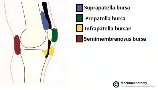
(1) Suprapatellar bursa: quadriceps femoris와 femur 사이
(2) Prepatellar bursa: patella 앞 피부 밑
(3) Infrapatellar bursa: patellar ligament의 전후에 위치 (deep/superficial)
(4) Semimembranosus bursa: semimembranosus와 gastrocnemius 사이
5) 인대
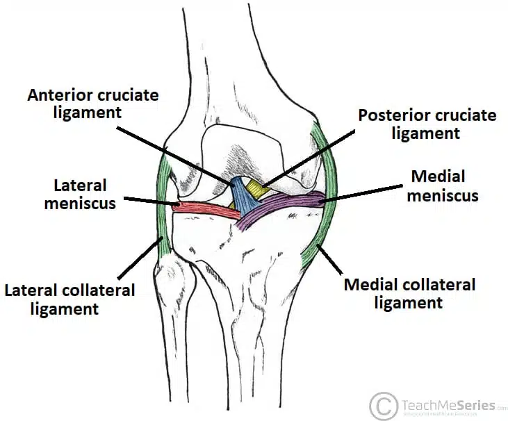
(!) Patellar ligament: quadriceps tendon의 연장으로 tibial tuberosity에 부착
(2) Medial collateral ligament (MCL): femoral medial epicondyle ↔ tibial medial condyle
(3) Lateral collateral ligament (LCL): femoral lateral epicondyle ↔ fibular head
(4) 십자인대: Anterior와 Posterior로 나뉨.
① Anterior cruciate ligament (ACL): tibial anterior intercondylar area ↔ femoral intercondylar fossa
→ tibia의 전방 탈구 방지
② Posterior cruciate ligament (PCL): tibial posterior intercondylar area ↔ femoral anteromedial condyle
→ tibia의 후방 탈구 방지
6) 임상적으로 중요한 포인트
(1) Collateral ligament injury
① Medial rotation 시 통증 → MCL 손상
② Lateral rotation 시 통증 → LCL 손상
(2) Cruciate ligament injury
① ACL 손상: 과신전 또는 무릎 굴곡 중 후방 충격 → anterior drawer test 양성
② PCL 손상: ‘dashboard injury’ (차 사고), 무릎 굴곡 상태에서 정강이 뒤로 밀림 → posterior drawer test 양성
(3) Bursitis
(4) Unhappy triad: MCL + medial meniscus + ACL 손상 동반. 주로 측방 충격에 의해 발생한다.
1. 종아리 개요
이론부터 문제까지, 알렌의 서재를 100% 활용하세요
※ 로그인 후 이용권 구매 시 전체 이용 가능합니다.
6,000개 이상의 문제와 연결되는 이론으로 개념과 적용을 한 번에
실제 국시와 동일한 CBT 환경으로 실전 감각 완성
틀린 문제를 매일 자동으로 챙겨주는 ‘오늘의 문제’
메모·암기카드·노트로 만드는 나만의 복습노트