발목과 발
이론과 하이라이트 히스토리를 확인 할 수 있어요.
0. 발목
항목 | 내용 |
|---|---|
관절 종류 | Synovial hinge joint |
관절 구성 | Tibia + Fibula + Talus (Calcaneus는 불포함) |
주요 움직임 | Dorsiflexion & Plantarflexion |
1) 관절면: Tibia + Fibula로 구성.
강한 tibiofibular ligaments로 고정되어 Mortise(절구) 구조 형성
2) 인대 구조
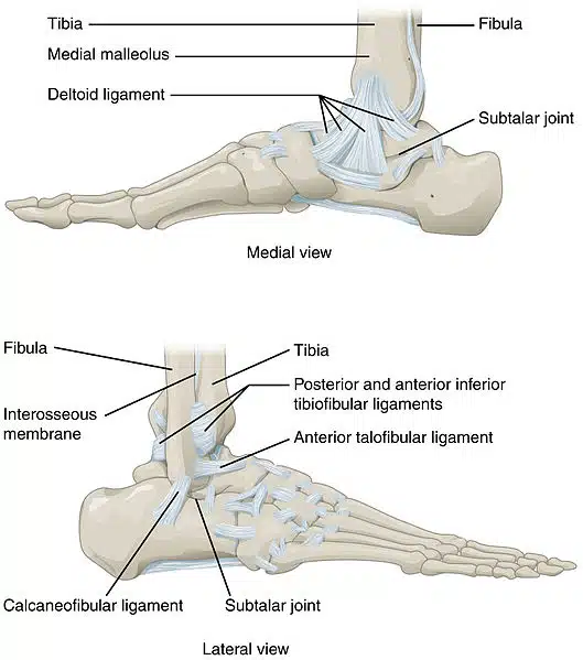
Website: TeachMeAnalysis
(1) Medial Ligament (Deltoid Ligament)
① Medial malleolus에서 일어 Talus, Calcaneus, Navicular 에 부착
② 기능: Eversion 저항
③ 구성: 4개 인대 (fan-shaped)
(2) Lateral Ligament
① Lateral malleolus에서 일어나 Talus 등으로 주행한다.
② 기능: Inversion 저항
③ 구성:
• Anterior talofibular lig.
• Posterior talofibular lig.
• Calcaneofibular lig.
• Ankle Sprain(발목 염좌)은 무게가 실린 상태에서 과도한 inversion + plantarflexion에 의해 일어난다.
- 가장 취약한 인대: Anterior talofibular lig. (Lateral lig.가 medial보다 약함 + inversion 시 긴장됨)
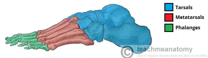
1. 발의 뼈
구분 | 구성 |
|---|---|
Tarsals | 발목 부위의 7개 불규칙 모양 뼈 |
Metatarsals | Tarsals와 Phalanges를 연결하는 5개의 뼈 |
Phalanges | 발가락 뼈 (2~5번째 발가락: 3개씩 / 엄지발가락: 2개) |
1) Tarsals
구분 | 뼈 | 특징 |
|---|---|---|
Proximal | Talus | 몸무게 전달, Ankle joint 형성 (Tibia, Fibula와 관절) |
Calcaneus | Heel bone, Achilles tendon 부착 | |
Intermediate | Navicular | Medial 위치, Tibialis posterior tendon 부착 |
Distal | Cuboid, Cuneiforms (Medial, Intermediate, Lateral) | Cuboid: Fibularis longus tendon 통과 |
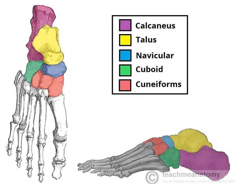
(1) 임상적으로 중요한 뼈: Talus, Calcaneus
• Talus fracture: Neck 부위 골절 흔함. 무혈성괴사(Avascular necrosis) 위험.
• Calcaneus fracture: 고에너지 축방향 외상 (낙상 등) → 아래목말관절(Subtalar joint) 손상 및 이후 관절염 가능성 있음.
2) Metatarsals
항목 | 설명 |
|---|---|
구조 | Base, Shaft, Neck, Head로 구성 |
관절 | Tarsometatarsal, Intermetatarsal, Metatarsophalangeal joints |
특징 | 1번~5번까지 번호 부여 (Medial → Lateral 방향) |
3) Phalanges
항목 | 설명 |
|---|---|
구성 | Base, Shaft, Head |
발가락별 수 | 2~5번 발가락: Proximal, Middle, Distal / 1번 발가락(엄지): Proximal, Distal |
2. 발의 근육
: 발의 근육은 종아리에서 오는 Extrinsic muscle과 발 내의 Intrinsic muscle로 구분할 수 있다.
Intrinsic muscle은 발등(Dorsal)쪽과 발바닥(Plantar)쪽으로 다시 구분된다.
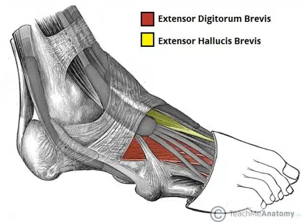
1) Dorsal muscles
Muscle | Attachments | Actions | Innervation |
|---|---|---|---|
Extensor digitorum brevis | Calcaneus → Long extensor tendons (2-4번째 발가락) | 2-4번째 발가락 extension | Deep fibular n. |
Extensor hallucis brevis | Calcaneus → Base of proximal phalanx of great toe | 엄지 extension |
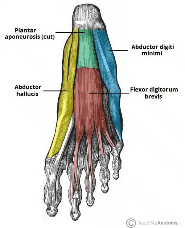
2) Plantar muscles
: 총 10개 근육이 4개 층으로 구분된다.
Tibial nerve의 분지인 Medial & Lateral plantar n.가 담당한다.
(1) 1st layer (발바닥에 가장 가까운 쪽)
근육 | 운동 | 신경 |
|---|---|---|
Abductor hallucis | 엄지발가락 abduction & flexion | Medial plantar n. |
Flexor digitorum brevis | Flexion of lateral 4 toes (PIP joints) | |
Abductor digiti minimi | 새끼발가락 abduction & flexion | Lateral plantar nerve |
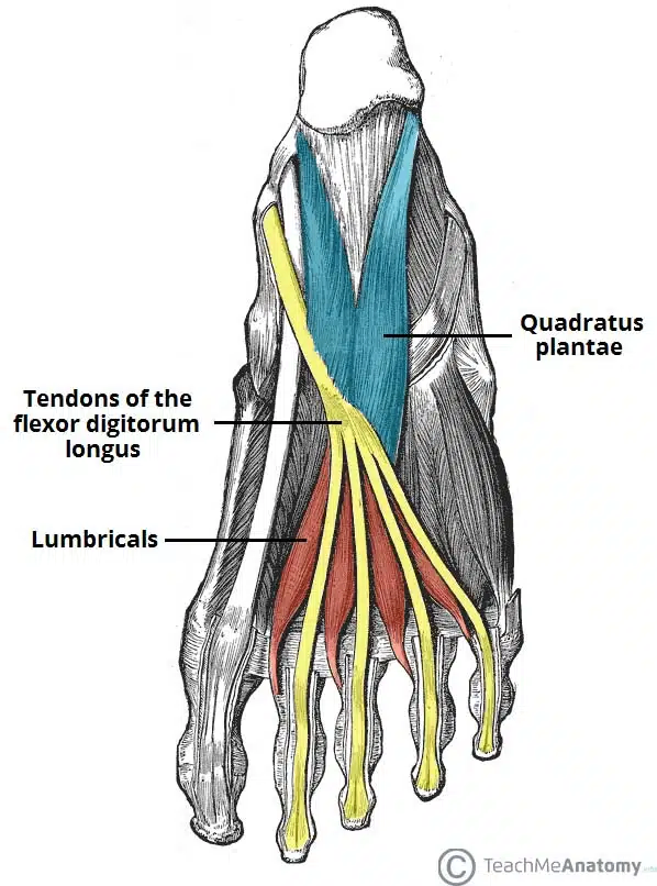
(2) 2nd layer
근육 | 운동 | 신경 |
|---|---|---|
Quadratus plantae | 발가락 flexion에서 F. digitorum longus 보조 | Lateral plantar n. |
Lumbricals (4) | 발허리발가락관절(MTP) flexion, | 1st: Medial plantar n. 2–4th: Lateral plantar n. |
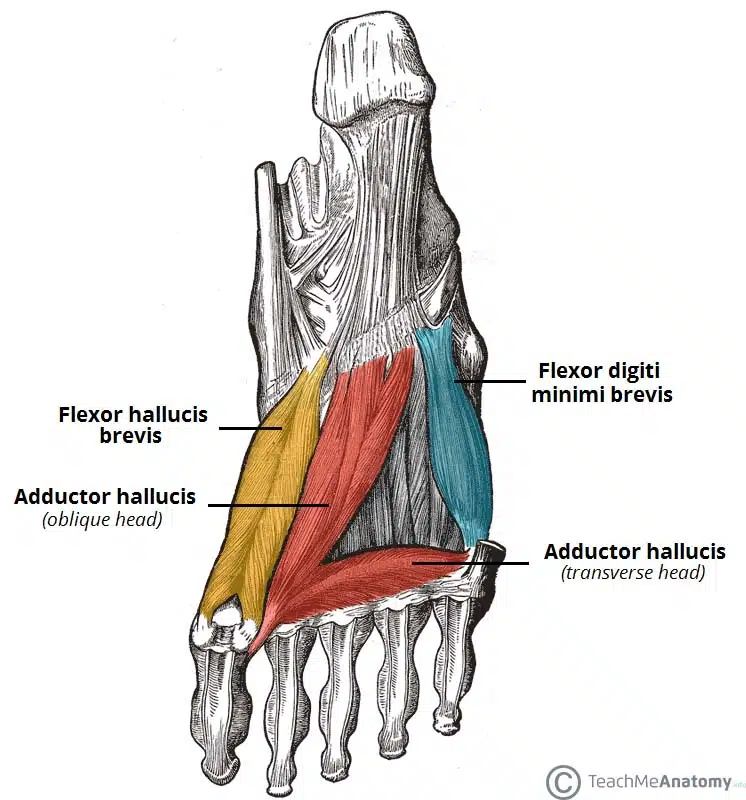
(3) 3rd layer
Muscle | Action | Innervation |
|---|---|---|
Flexor hallucis brevis | 엄지발가락 flexion (MTP joint) | Medial plantar n. |
Adductor hallucis (Oblique + Transverse head) | 엄지발가락 adduction, transverse arch support | Deep branch of lateral plantar n. |
Flexor digiti minimi brevis | 새끼발가락 flexion (MTP joint) | Superficial branch of lateral plantar n. |
(4) 4th layer
근육 | 운동 | 신경 |
|---|---|---|
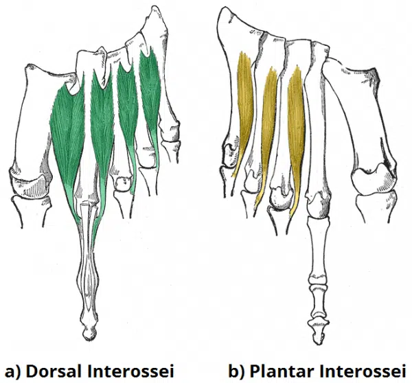
Plantar interossei (3) | Adduction of digits 3–5, MTP flexion | Lateral plantar n. |
Dorsal interossei (4) | Abduction of digits 2–4, MTP flexion |
형태: Plantar interossei → Unipennate (한쪽 깃모양) / Dorsal interossei → Bipennate (양쪽 깃모양)