심장
이론과 하이라이트 히스토리를 확인 할 수 있어요.
심장과 관련된 기본적인 지식(구조 및 특징, 관상동맥 등)은 외우고 있어야 한다.
특히 심장에서 확인하고 싶은 부위별 청진 위치가 자주 출제되니 꼭 알아놓자.
0. 심장 구조
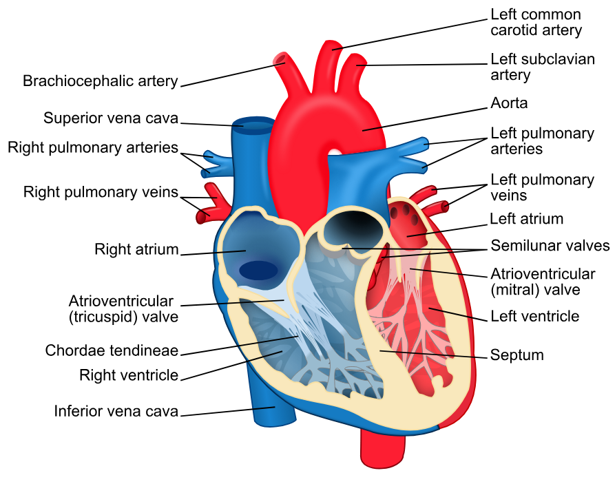
1. 심장의 큰 혈관들
혈관 | 기원 | 경로 | 설명 |
|---|---|---|---|
Aorta | 좌심실 (Left ventricle) | Ascending aorta → Arch of aorta → Descending aorta | 동맥혈을 전신으로 운반하는 가장 큰 동맥. 관상동맥은 ascending aorta에서 분지. |
Pulmonary arteries | 우심실 (Right ventricle) | Pulmonary trunk → Right and Left pulmonary arteries | 정맥혈을 폐로 운반. |
Pulmonary veins | 폐 (Lungs) | 우/좌, 상/하 폐정맥 4개 | 동맥혈을 좌심방으로 운반. |
Superior vena cava (SVC) | 좌우 Brachiocephalic vein 합류 | 직접 우심방으로 유입 | 상체에서 발생한 정맥혈을 심장으로 운반. |
Inferior vena cava (IVC) | 좌우 Common iliac vein 합류 | T8 수준에서 횡격막을 통과하여 우심방으로 유입 | 하체에서 발생한 정맥혈을 심장으로 운반. |
2. 심방과 심실
이론부터 문제까지, 알렌의 서재를 100% 활용하세요
※ 로그인 후 이용권 구매 시 전체 이용 가능합니다.
6,000개 이상의 문제와 연결되는 이론으로 개념과 적용을 한 번에
실제 국시와 동일한 CBT 환경으로 실전 감각 완성
틀린 문제를 매일 자동으로 챙겨주는 ‘오늘의 문제’
메모·암기카드·노트로 만드는 나만의 복습노트
커뮤니티 Q&A
위 이론과 관련된 게시글이에요.
이해가 안 되거나 궁금한 점이 있다면 커뮤니티에 질문해 보세요!
로그인