위샘의 조직학
이론과 하이라이트 히스토리를 확인 할 수 있어요.
위샘의 조직학
•위샘은 점막층(mucosa)내에 위치하며 표면상피로 덮여 있음 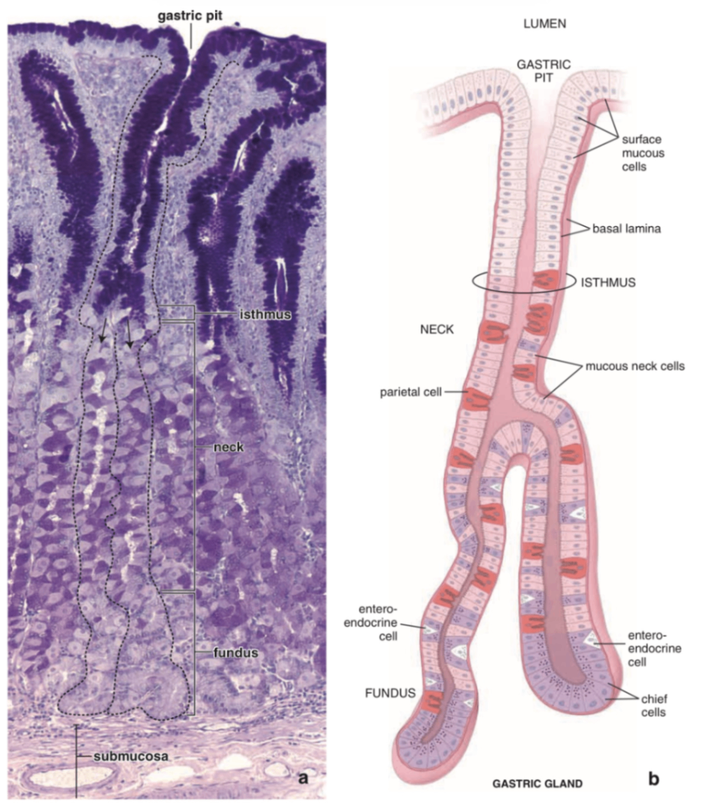
세포 | 위치 | 형태학적 특징 | 기능 |
|---|---|---|---|
표면상피세포(surface mucous cell) | 위소와를 덮는 상피 | 단일 원주상피, 점액 포함 | 알칼리성 점액 분비 → 위산(HCl)으로부터 상피 보호 |
목 점액세포(mucous neck cell) | 샘의 neck 부위 | 점액 과립, 핵은 basally located | 점액 분비 |
벽세포(parietal cell) | neck ~ fundus 부위 | 크고 eosinophilic, 둥근 핵, 내재성 위관(intracellular canaliculi) | HCl 분비, 내인자(intrinsic factor) 분비 (Vit B12 흡수에 필수) |
주세포(chief cell) | fundus | basophilic (rER 풍부), apical granules | 펩시노겐 분비 → 위산에 의해 펩신으로 활성화 |
내분비세포(enteroendocrine cell) | fundus | 기저부에 위치, H&E에선 구분 어려움(Ag/Cr 염색에서 확인 가능) | • G세포: gastrin 분비 → 벽세포 자극 • D세포: somatostatin, serotonin, histamine 등 분비 |
이론부터 문제까지, 알렌의 서재를 100% 활용하세요
※ 로그인 후 이용권 구매 시 전체 이용 가능합니다.
6,000개 이상의 문제와 연결되는 이론으로 개념과 적용을 한 번에
실제 국시와 동일한 CBT 환경으로 실전 감각 완성
틀린 문제를 매일 자동으로 챙겨주는 ‘오늘의 문제’
메모·암기카드·노트로 만드는 나만의 복습노트
커뮤니티 Q&A
위 이론과 관련된 게시글이에요.
이해가 안 되거나 궁금한 점이 있다면 커뮤니티에 질문해 보세요!
로그인